CASE STUDY: Recurrent Rectal Prolapse in a Manx Cat: A Rare Congenital Cystic Anomaly Resolved by Transanal Surgery
Patients' Stories

Recurrent Rectal Prolapse in a Manx Cat: A Rare Congenital Cystic Anomaly Resolved by Transanal Surgery
Animal Emergency & Referral Associates (AERA) | June 2026
BACKGROUND AND PATIENT HISTORY
Patient: Veerkat
Signalment: 7-year-old female spayed Manx
Primary Complaint: Recurrent rectal prolapse and dyschezia
Chronicity: Episodic, approximately 1–2 times per year since early in life
Surgeon: Brian Brophy, MVB
Care Coordinator: Joanne Zimmermann, CVT, VTS (Anesthesia/Analgesia)
Outcome: Symptom-free at 18+ months post-operatively
At five months old, Veerkat was surrendered to AERA after a treating emergency clinician, declining to perform euthanasia for a presumed rectal prolapse, recognized that further evaluation was warranted. Joanne Zimmermann, CVT, VTS (Anesthesia/Analgesia) in the AERA Surgery Department since May 2000, took Veerkat into her personal care and managed her condition symptomatically over the following years.
Despite diligent supportive management, Veerkat continued to experience episodic rectal prolapse. Joanne's persistence and decision to pursue advanced imaging set the stage for a definitive diagnosis and cure.
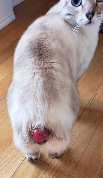
CLINICAL PRESENTATION
Dr. Brian Brophy, MVB, assumed primary oversight of Veerkat's surgical care in February 2025. At that time, her clinical pattern was well-established: recurrent episodes of rectal prolapse and dyschezia occurring one to two times annually. Each episode was accompanied by a palpable swelling or mass effect dorsal to the rectum: approximately 3 cm in diameter, slightly firm, fluid-filled and turgid.
During these episodes, palliative management consisted of a small stab incision into the affected area with manual aspiration of the accumulated fluid. The fluid was consistently thick, viscous and opaque tan in color. Serial cultures were uniformly negative, ruling out an infectious etiology. Symptom resolution following aspiration was temporary, and recurrence was reliable.
DIAGNOSTIC WORKUP
Given the chronicity and pattern of recurrence, Dr. Brophy recommended advanced cross-sectional imaging to characterize the lesion prior to any definitive surgical planning. Joanne ultimately agreed, and CT of the pelvis and perineum was performed.
CT Findings: Imaging revealed a blind-ending, focal, fluid-filled structure situated dorsal to the rectum and left of the distal rectal/anal junction. The lesion was causing focal compression of the rectum with proximal dilation. The attenuation of the contents was higher than expected for simple fluid, raising suspicion for inspissated material, a proteinaceous abscess, or impacted fecal/fluid material. The structure did not demonstrate significant contrast enhancement.
The radiologist's interpretation noted the impossibility of clearly differentiating the lesion margins from the rectal wall, raising concern for resectability and a potentially high risk of post-operative fecal incontinence. The differential at that time included anal sac origin, rectal diverticulum, or another cystic anomaly. Ultrasound was recommended as a potential adjunct, though surgery ultimately proceeded on the basis of the CT and the patient's clinical history.
SURGICAL INTERVENTION
Dr. Brophy initially approached the lesion via a dorsal perineal approach. Intraoperatively, however, the external surface of the rectal wall in this region appeared grossly normal, with no discrete lesion identifiable from the outside. The dorsal approach was closed, and the surgical strategy was revised.
A transanal approach was then employed: the rectum was everted, and the cystic structure was brought into the operative field. Upon incision and drainage of the fluid, the interior lining of the structure was found to be composed of rectal mucosal tissue: clearly distinct from the surrounding normal rectal mucosa and entirely non-communicating with the rectal lumen. This finding was consistent with a congenital duplication cyst or vestigial cystic anomaly of the rectum.
All aberrant mucosal tissue was meticulously dissected from the cystic wall, and the resulting defect in the rectal wall was closed primarily. Tissue was submitted for histopathology.
OUTCOME AND FOLLOW-UP
Veerkat's recovery was uneventful. Since surgery, she has experienced no recurrence of perirectal swelling, rectal prolapse, or dyschezia — now approximately 18 months post-operatively.
CLINICAL TAKEAWAYS
This case is a compelling example of a rare congenital rectal anomaly presenting as what appeared to be a straightforward, if persistent, rectal prolapse. Several features of Veerkat's presentation are instructive:
1. Chronicity and pattern recognition matter. Recurrent, culturally negative perirectal fluid accumulation with a palpable dorsal mass is not a typical prolapse presentation. Recognizing this pattern over time was the impetus for advanced imaging.
2. CT is essential for complex perirectal pathology. Cross-sectional imaging defined the location, character and relationships of the lesion — information unavailable on physical examination alone.
3. Surgical adaptability is critical. When the dorsal approach did not reveal the expected anatomy, pivoting to a transanal technique allowed direct access and ultimately curative resection.
4. Histopathology confirms and educates. Submission of the tissue for pathologic analysis provided definitive characterization of the anomalous mucosal lining.
Rectal duplication cysts are exceedingly rare in companion animals. This case adds to the small body of literature on cystic perirectal anomalies in cats and demonstrates that long-standing, seemingly refractory colorectal disease may harbor an anatomic etiology amenable to surgical cure.